Endobronchial Ultrasound (EBUS)
Endobronchial ultrasound, also known as EBUS, is a minimally invasive technique is helpful in the diagnosis of lung cancer, lymphomas, infections and other diseases causing enlarged lymph nodes and tumors in the chest.
It allows physicians to display real-time, improved images and gives them the ability to easily view difficult-to-reach areas of your respiratory system.
Benefits of EBUS
Benefits of EBUS include:
- Higher accuracy
- No required cutting or incisions
- The opportunity for immediate surgery if necessary
- Ability for patients to go home the same day if no further surgery is required
What to Expect
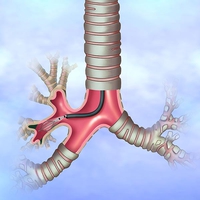
During the procedure, a flexible tube (called a bronchoscope) is inserted into the windpipe (trachea).
Using a camera and ultrasound technology, the scope obtains superior images of your lungs and nearby lymph nodes.
Your pulmonologist also will perform a technique known as a transbronchial needle aspiration (TBNA) to obtain tissue and fluid samples from the lungs and surrounding lymph nodes.
These will be used to perform a biopsy.
You will be sedated during the procedure. Risks include possible bleeding and infection.
Although EBUS usually is an outpatient procedure, complications may result in a brief hospital stay.